


Why Is a Doppler Ultrasound Pregnancy in Third Trimester Important?
The nine months of pregnancy are filled with excitement, emotions, and many physical changes. During this journey, regular medical checkups become very important to monitor the health and growth of the baby. Ultrasound scans are one of the most essential tests recommended by doctors during pregnancy.
Pregnancy is divided into three stages:
Many women often ask, “Doppler ultrasound pregnancy in which month is done?” or “Doppler test in pregnancy in which month is most important?” Usually, a Doppler ultrasound pregnancy in third trimester is advised because this is the phase when the baby grows rapidly and prepares for birth.
What Is Doppler Ultrasound Pregnancy in Third Trimester?
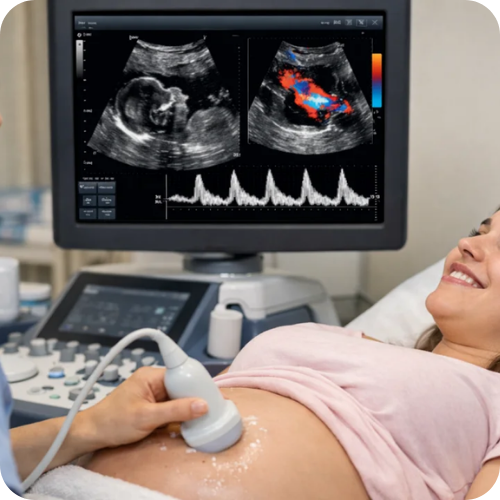
A Doppler ultrasound is a specialised ultrasound test that helps doctors examine the blood flow through the placenta and umbilical cord. It is commonly recommended for women who have high blood pressure during pregnancy or when doctors suspect restricted growth of the baby inside the womb.
When patients ask, “Doppler scan in which month should be done?” doctors generally recommend it during the last three months of pregnancy. The scan helps gynaecologists study the blood vessels, arteries, veins, and the overall condition of the baby.
Doppler Test in Pregnancy in Which Month Is Recommended?
One of the most common questions asked by expecting mothers is, “Doppler ultrasound pregnancy in which month is necessary?” The answer depends on the pregnancy condition. However, in most cases, doctors suggest a Doppler ultrasound pregnancy in third trimester, especially after 28 weeks.
The final trimester is considered very important because the baby reaches full development during this stage. The baby also changes position inside the womb while getting ready for delivery. Monitoring blood circulation during this period becomes essential.
A Doppler test in pregnancy in which month becomes more critical when there are complications like:
- High blood pressure during pregnancy
- Diabetes
- Reduced baby movements
- Suspected growth restriction
- Placenta-related concerns
Why Is Doppler Ultrasound Pregnancy in Third Trimester So Important?
The last three months of pregnancy are crucial for both the mother and the baby. During this period, the fetus grows quickly, gains weight, and develops stronger muscles.
A Doppler ultrasound pregnancy in third trimester helps doctors check whether the baby is receiving enough oxygen and nutrients through proper blood circulation. It can also detect reduced blood flow in the umbilical artery.
Blood flow is extremely important because oxygen reaches the baby through the bloodstream. If the blood supply is interrupted, it may affect the baby’s growth and overall health.
This is why doctors may recommend repeated scans in the third trimester to monitor the condition closely.
Doppler Scan in Which Month Helps Detect Delivery Position?
Many women ask, “Doppler scan in which month helps identify delivery type?” During the final trimester, the Doppler ultrasound can help doctors understand the baby’s position inside the womb.
If the baby’s head is facing downward, there are higher chances of a normal vaginal delivery. However, if the baby remains in a breech position, doctors may recommend a caesarean section.
The scan also helps evaluate:
- Placenta position
- Placenta maturity
- Cervix condition
- Amniotic fluid levels
Doctors ensure that the placenta does not block the cervix opening, which is important for safe delivery.
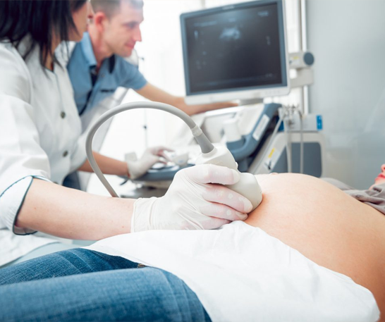
How Is Doppler Ultrasound Done During Pregnancy?
A Doppler ultrasound pregnancy in third trimester is usually performed using an abdominal probe. By this stage, the baby’s organs are fully developed, and the foetus appears more like a new-born infant.
During the scan, sound waves create images of the baby and measure blood circulation. The procedure is painless and safe for both mother and baby.
The scan allows doctors to monitor:
- Umbilical cord blood flow
- Placental circulation
- Baby’s heartbeat
- Fetal growth and movements
Why Repeated Doppler Tests May Be Needed
Sometimes doctors may advise multiple Doppler scans during the final months of pregnancy. This usually happens when the baby needs close monitoring.
If reduced oxygen supply or restricted blood flow is detected, doctors may consider early delivery to protect the baby’s health.
Many expecting mothers wonder, “Doppler test in pregnancy in which month should be repeated?” The answer depends on the baby’s condition and the mother’s health. In high-risk pregnancies, repeated monitoring during the third trimester becomes necessary.
Doppler Ultrasound Helps Estimate Baby’s Growth
Another important benefit of Doppler ultrasound pregnancy in third trimester is that it helps doctors estimate the baby’s birth weight and growth pattern.
Measurements are taken from:
- Skull size
- Abdomen diameter
- Thigh bone length
These details help doctors understand whether the baby is developing normally.
A Doppler ultrasound gives doctors critical information before delivery. It helps assess the baby’s growth, oxygen supply, placenta condition, and delivery position. This allows doctors and mothers to prepare better for childbirth, whether it is a normal delivery (NVD) or a caesarean section (LSCS).
![]()
CRH ENT, MRI & Diagnostics brings to Delhi NCR a unique concept of a dedicated single speciality ENT centre and the complete range of diagnostic services under one roof.


